Edutek Instrumentations Laboratory Chemicals

- Products
- Microscope-Equipments
- Fluorescent Microscope
Product Range
Fact Sheet
- Location:Haryana, India
- Year of Establishment:2009
- Business Type:Manufacturer, Exporter
- Turnover:Rs. 50 Lakh - 5 Crore
(or USD 100 K - 1 Million) - Main Products:Laboratory Instrument, Biology Equipment
- Reviews & Rating:

Get Verified, Sell more with
- Buyer's trust
- Faster conversions
- Better Rankings
- More
Its Free
Verify NowFluorescent Microscope
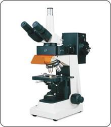
Fluorescent Microscope is used for capillary examination. This technique is a new, highly sensitive rapid method of diagnosing malaria.
- FOB PriceNA
- Min Order QuantityNA
- Payment TermsNA

Other Details
Fluorescent Microscope is used for capillary examination. This technique is a new, highly sensitive rapid method of diagnosing malaria. It is also useful in the diagnosis of filariasis and leptospirosis. The principle of this method is the malaria parasite picks up fluorescent stain into their nucleus and cytoplasm, so that its morphologic characteristics can be examined by fluorescent microscopy. The nucleus appears green and the cytoplasm reddish orange,
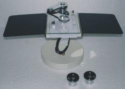
The Capillary is coated with Acridine orange on its inside. Parents Blood is loaded into this capillary and centrifuged at 12000 rpm for5 minutes. The Blood components settle at different levels in the capillary. These different layers are displayed in the picture.
Examination of the area just below granulocyte-RBC junction helps to detect the malarial parasites, as these are concentrated in this are upto 1000 times. Examination of Lymphocyte / monocyte layer using transmitted light helps to detect the schizonts, and gametocytes which gets concentrated in this layer very easily.
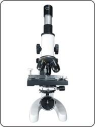
Method of Examination:
The granulocyte layer is then seen under 62x oil objective. By changing the light source to fluorescent light the parasitic forms can be seen as greenish orange stained structures. Using only transmitted light the parasitic forms are seen as block pigmented structures as shown in the preceding photomicrographs.
Other Information
Packaging Details: This microscope we prefer for the personal delivery and the demonstration but still if it is not possible then it is securely packed in two different thermocol boxes.
Images